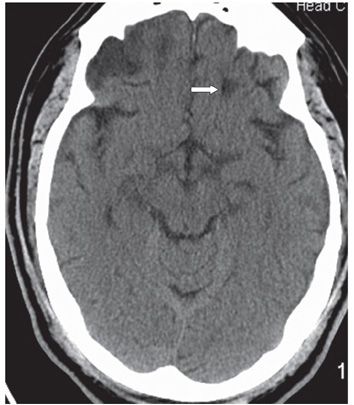
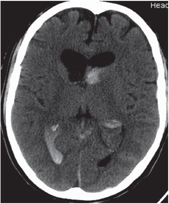
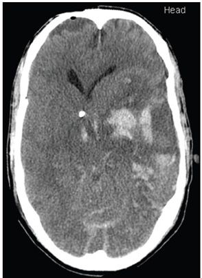
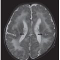
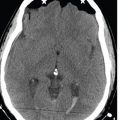
FINDINGS Figure 57-1. Axial NCCT through inferior frontal lobes. There is a 1.5-cm round hyperdensity surrounded by a hypodense halo in the left frontal lobe just posterior and superior to the left orbital roof (arrow) consistent with hematoma. There are contusions in the right frontal and left temporal lobes. Figure 57-2. Axial NCCT through the frontal lobes 6 months after Figure 57-1. The area of hematoma is now represented by a well-defined hypodensity (encephalomalacia) significantly smaller than the original lesion (arrow). Figures 57-3 and 57-4. Axial NCCT 24 hours apart in another patient. There are new and expanding hematomas (arrows in Figure 57-4) on follow-up. There is significant brain swelling, mass effect, and herniation.
DIFFERENTIAL DIAGNOSIS N/A.
DIAGNOSIS Traumatic intracerebral hematoma (TICH).
DISCUSSION


Stay updated, free articles. Join our Telegram channel

Full access? Get Clinical Tree


