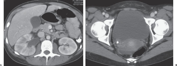
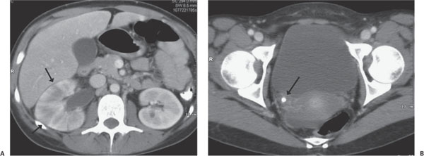
Case 57 A teenager with high fever and flank pain. (A,B) Axial computed tomography images at the level of the kidneys and urinary bladder after oral and intravenous contrast demonstrate a swollen right kidney with wedge-shaped areas of parenchymal hypoattenuation (“striated nephrogram,” arrow) and hydronephrosis. In addition, a several-millimeter calcific density is seen at the right ureterovesical junction (UVJ, arrow). • Obstructive uropathy:
Clinical Presentation
Imaging Findings
Differential Diagnosis
![]()
Stay updated, free articles. Join our Telegram channel

Full access? Get Clinical Tree


