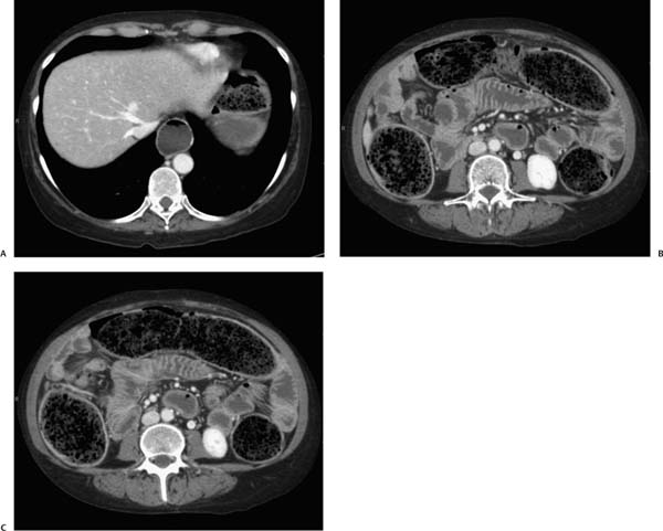
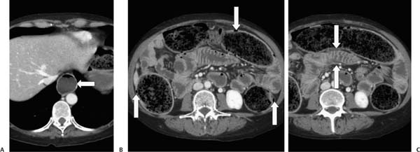
Case 57 A 51-year-old woman presents with constipation. (A) Contrast-enhanced computed tomography (CT) shows marked dilatation of the esophagus (arrow). (B) Marked retention of fecal debris is noted throughout the colon, and gas is present within the colon wall (arrows) without associated mural thickening or mesenteric fat stranding, consistent with pneumatosis cystoides coli. (C) Stacks of thin, straight folds within the jejunum (arrows) are noted. • Scleroderma: This is the diagnosis and is indicated by esophageal dilatation, marked fecal retention, the history of constipation, pneumatosis cystoides coli, and hide-bound small-bowel folds.
 Clinical Presentation
 Imaging Findings
 Differential Diagnosis
Stay updated, free articles. Join our Telegram channel

Full access? Get Clinical Tree


