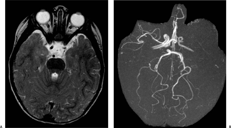
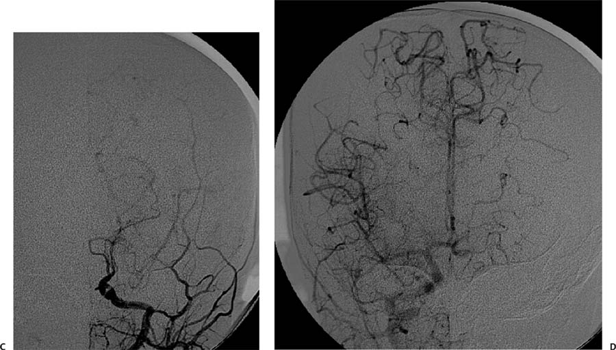
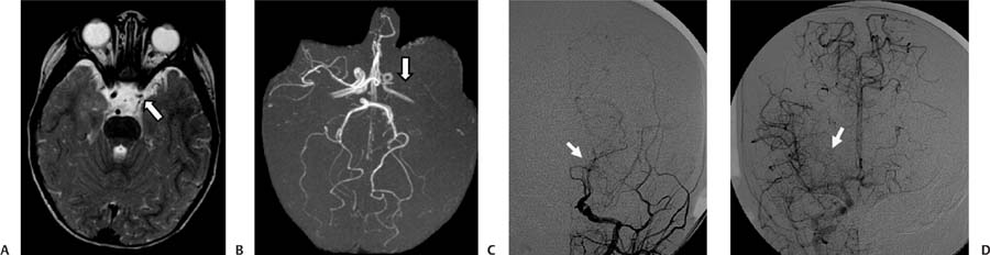
Case 58 A 19-month-old boy with the diagnosis of neurofibromatosis type 1 undergoing screening magnetic resonance imaging of the brain. (A) Axial T2-weighted image (WI) of the brain shows narrowing of the left internal carotid artery (ICA) in the supraclinoid segment (arrow). (B) Magnetic resonance angiography (MRA) of the brain reveals narrowing of the left ICA with an absent flow-related signal in the left middle cerebral artery (MCA; arrow). There is also a paucity of branches of the right MCA. (C) Digital subtraction angiogram (DSA) of the left common carotid artery shows tapering and occlusion of the distal ICA. Prominent leptomeningeal collateral vessels are noted (arrow). (D)
Clinical Presentation
Further Work-up
Imaging Findings
![]()
Stay updated, free articles. Join our Telegram channel

Full access? Get Clinical Tree


