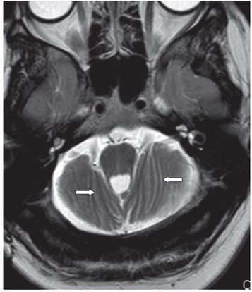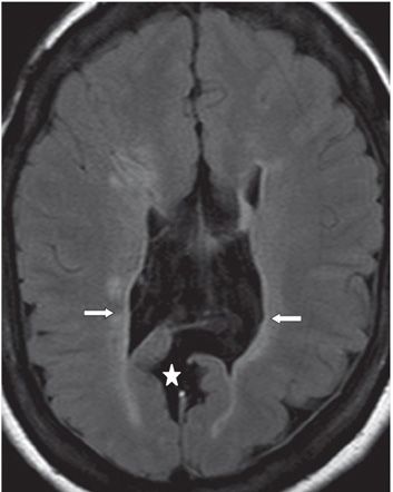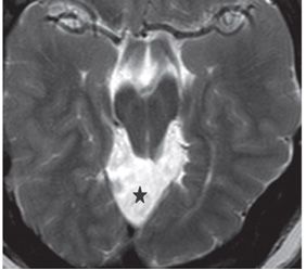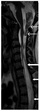


FINDINGS Figure 58-1. Sagittal MR T1WI. There is a very small posterior fossa with a very small cerebellum extending through wide foramen magnum into the upper cervical spinal canal (transverse thick arrows). The vertical thin arrow points to the low position of the torcula. There is a pointed (beaked) tectum (thin transverse arrow). The fourth ventricle is tubular and continuous inferiorly into the upper spinal canal with no visible fastigium (star). The body/splenium of the corpus callosum is severely atrophic (chevron). Parasagittal cerebral dysgenesis is present (right left arrow). The massa intermedia (heart) is large. Figure 58-2. Axial T2WI through the small posterior fossa. There is anteroposterior orientation of the cerebellar folia (transverse arrows). There is concavity of posterior petrosal surface. Figure 58-3. Axial FLAIR through the lateral ventricles. There is dysgenesis of the parasagittal occipital lobes and dilation of the posterior interhemispheric fissure (star). There is scalloping of the ventricular walls with periventricular hyperintensities due to leukomalacia (transverse arrows). There is bilateral cortical malformations, a mixture of pachygyria and polymicrogyria. Figure 58-4. Axial T2WI through midbrain. Beaked tectum protrudes into dilated quadrigeminal cistern (star). Figure 58-5
Stay updated, free articles. Join our Telegram channel

Full access? Get Clinical Tree


