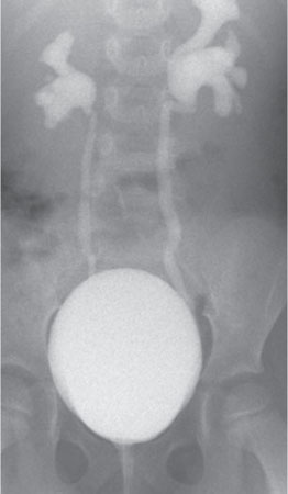
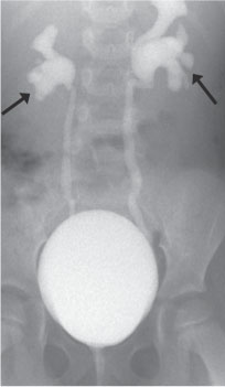
Case 58 A child with a recent history of urinary tract infection. A radiograph from a voiding cystourethrogram (VCUG) demonstrates bilateral vesicoureteral reflux during voiding (arrows). • Vesicoureteral reflux: Assuming that an initial scout radio-graph of the abdomen demonstrated no contrast material in the abdomen, these findings are diagnostic of vesicoureteral reflux. This degree of calyceal blunting and ureteral dilatation is consistent with grade 3 reflux. • Normal bowel or bone anatomy: Depending on the distribution of bowel gas and the degree of obliquity of the images, normal anatomy can sometimes mimic a contrast-opacified ureter, although that would not be a reasonable hypothesis in this case. • Prior administration of contrast: Inexperienced observers sometimes mistakenly interpret the radiographic findings of patients who have received intravenous contrast material as vesicoureteral reflux. In general, there will be opacification of the renal parenchyma in such cases, and it would be rare for both ureters to be as well opacified as in this case.
Clinical Presentation
Imaging Findings
Differential Diagnosis
Essential Facts
Stay updated, free articles. Join our Telegram channel

Full access? Get Clinical Tree


