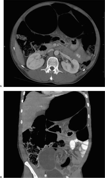
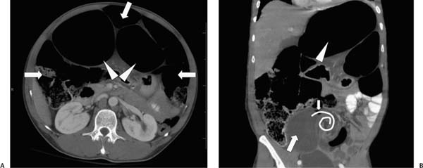
Case 58 A 43-year-old man presents with intense lower abdominal pain and distension. (A) Contrast-enhanced computed tomography (CT) shows two markedly dilated bowel segments (arrowheads) in the midabdomen surrounded by nondilated segments of ascending, transverse, and descending colon (arrows). (B) Coronal reformatted image shows a markedly dilated segment of bowel (arrowhead) with adjacent, nondilated transverse colon. A dilated, fluid-filled bowel segment (large arrow) in the lower abdomen terminates with a beaklike configuration (small arrow), followed by a whirl of mesenteric vessels and fat (curved line). • Sigmoid volvulus: This diagnosis is strongly indicated by the CT findings of the whirl sign, the beak sign, and a markedly distended colonic segment with nondilated ascending, transverse, and descending colon. • Adhesion:
 Clinical Presentation
 Imaging Findings
 Differential Diagnosis
![]()
Stay updated, free articles. Join our Telegram channel

Full access? Get Clinical Tree


