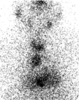
CASE 58 A 55-year-old woman presents with a large mediastinal mass on CT of the thorax. Thyroid function tests show normal values: thyroid-stimulating hormone (TSH), 0.1 μU/mL;, thyroxine (T4), 10.5 μg/dL. Fig. 58.1 Anterior parallel-hole collimator view of neck and thorax, 123I. • 0.500 mCi of 123I administered orally 4 hours before scan • Five-minute anterior parallel-hole collimator view (Fig. 58.1); SPECT with low-energy, high-resolution collimators on dual-detector gamma camera • Acquisition parameters • Processing parameters
Clinical Presentation
Technique
 64 × 64 matrix
 180 degrees
 64 views
 30 seconds per view per detector
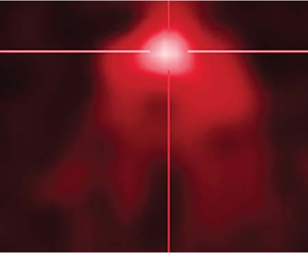
 Gaussian iterative reconstruction, filter 10.0 (Fig. 58.2)
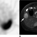
 Thoracic CT (Fig. 58.3)
 Fused SPECT/CT (Fig. 58.4) with commercial software
Related posts:


Stay updated, free articles. Join our Telegram channel

Full access? Get Clinical Tree


