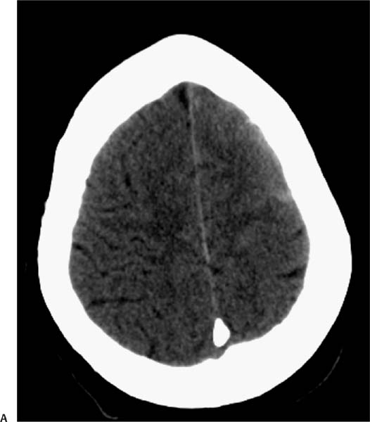
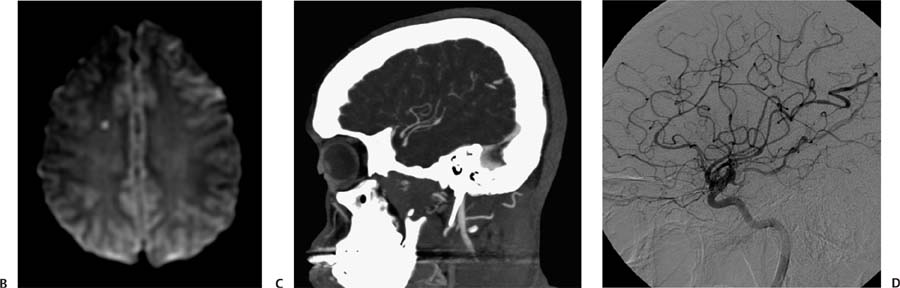
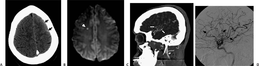
Case 59 A 24-year-old postpartum patient with a history of headache. (A) Nonenhanced computed tomography (CT) of the brain demonstrates subarachnoid hemorrhage in the left frontal convexity (arrows). (B) Diffusion-weighted image shows a tiny focus of acute ischemia in the right centrum semiovale (arrowhead). Fluid-attenuated inversion recovery images (not shown) confirmed subarachnoid hemorrhage in the left convexity. (C) CT angiography, sagittal maximum-intensity-projection image, reveals a beaded appearance of some of the left middle cerebral artery (MCA) branches. (D) Lateral projection of a digital subtraction angiogram of the left internal carotid artery (ICA) shows multiple areas in the anterior cerebral artery and MCA branches with alternating segments of narrowing and dilatation (i.e., beading; arrows). • Central nervous system vasculitis:
Clinical Presentation
Further Work-up
Imaging Findings
Differential Diagnosis
![]()
Stay updated, free articles. Join our Telegram channel

Full access? Get Clinical Tree


