 Clinical Presentation
A 50-year-old man with back pain.
Further Work-up
 Imaging Findings
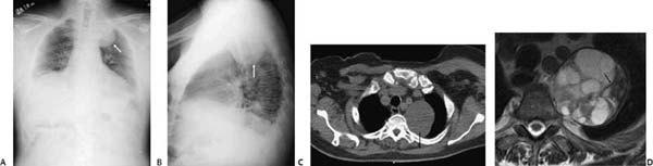
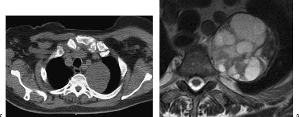
(A) Frontal chest radiograph demonstrates a large, well-circumscribed mass in the left upper chest (arrow). There is no evidence of rib destruction. (B) Lateral chest radiograph confirms the smooth inferior margin of the mass (arrow); however, the precise location of the abnormality cannot be determined. (C) Noncontrast computed tomography (soft-tissue windows) shows that the mass has heterogeneous density. It forms obtuse angles with the pleura, suggesting an extrapulmonary location (arrow). (D) T2-weighted magnetic resonance imaging (MRI) shows that the mass has heterogeneous but predominantly high signal intensity and multiple hypointense septa (arrow).
Stay updated, free articles. Join our Telegram channel

Full access? Get Clinical Tree


