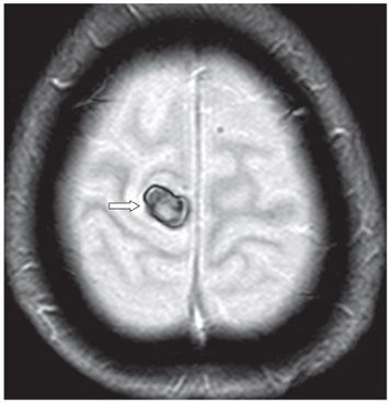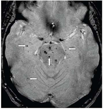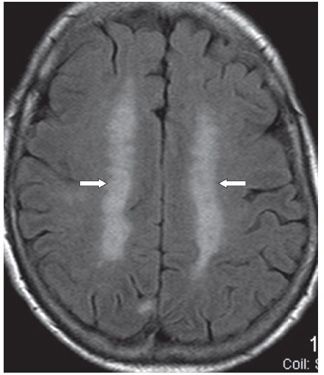


FINDINGS Figure 59-1. Axial NCCT through the vertex. There is a 1.5 cm × 1.2 cm subcortical right frontal parasagittal premotor well-defined hyperdensity with surrounding hypointense halo consistent with acute hemorrhage with surrounding edema (arrow). Figure 59-2. Axial MR GRE through the vertex. There is an ovoid hypointense (blooming) mass in the right frontal premotor cortex (arrow). Figure 59-3. Axial GRE through the upper pons. There are multifocal punctate hypointensities in the pons and bilateral cerebral hemispheres (arrows) consistent with microhemorrhages. Figure 59-4. Axial FLAIR through the centrum semiovale. There is bilateral almost symmetrical confluent WM hyperintensity (arrows).
DIFFERENTIAL DIAGNOSIS Cerebral cavernous malformation (CCM), hypertensive intracerebral hematoma (ICH), amyloid angiopathy-related ICH, hemorrhagic metastasis.
DIAGNOSIS Right frontal lobar hematoma with microhemorrhages.
DISCUSSION
Stay updated, free articles. Join our Telegram channel

Full access? Get Clinical Tree


