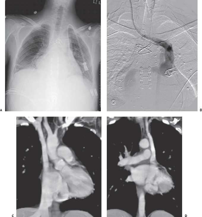
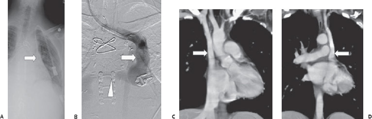
Case 59 A 41-year-old man presents for placement of a central line for dialysis. (A) Frontal radiograph shows central line along the left mediastinum (arrow). The line was not functioning. (B) Venogram from a new right jugular line shows a left-sided superior vena cava (SVC: arrow) with anomalous drainage into the coronary sinus (arrowhead). (C) Coronal reformatted contrast-enhanced computed tomographic (CT) scan shows patent right SVC (arrow). (D) More posterior slice shows persistent left SVC (arrow).
 Clinical Presentation
 Imaging Findings
 Differential Diagnosis
Stay updated, free articles. Join our Telegram channel

Full access? Get Clinical Tree


