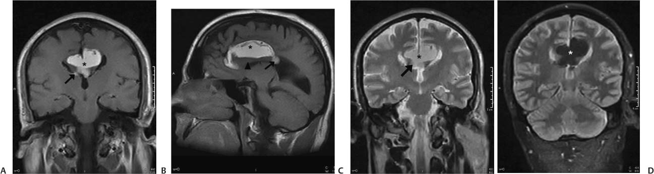
Case 6 A 30-year-old man with a history of headaches. (A) Coronal T1-weighted magnetic resonance imaging (MRI) of the brain demonstrates a high-signal lesion in the midline (asterisk) at the site of the corpus callosum. The lesion shows ventricular extension (arrow). (B) Sagittal T1-weighted MRI of the brain demonstrates a high-signal lesion in the midline (asterisk) that shows ventricular extension (arrow). Note the hypoplasia of the corpus callosum (arrowhead). (C) Coronal T2-weighted MRI of the brain demonstrates a high-signal lesion in the midline (asterisk) at the site of the corpus callosum. The lesion shows ventricular extension (arrow). (D) Coronal T2-weighted fat-saturated MRI of the brain demonstrates loss of signal in the midline lesion (asterisk), which was bright before fat saturation. • Intracranial lipoma:
Clinical Presentation
Imaging Findings
Differential Diagnosis
![]()
Stay updated, free articles. Join our Telegram channel

Full access? Get Clinical Tree


