 Clinical Presentation
A 57-year-old man with cough.
Further Work-up
 Imaging Findings
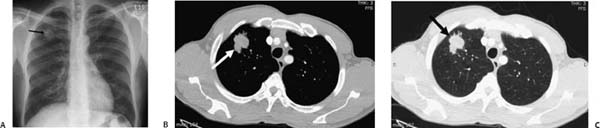
(A) Conventional radiograph of the chest reveals a lobulated, irregular 2.5-cm nodule in the right upper lobe (arrow). (B, C) Contrast-enhanced computed tomography (CT) images with mediastinal (B) and lung (C) windows confirm the presence of a spiculated, noncalcified, soft-tissue-density nodule in the anterior segment of the right upper lobe (arrows).
 Differential Diagnosis
• Lung cancer presenting as a solitary pulmonary nodule (SPN): A spiculated, noncalcified pulmonary nodule in an adult patient should be considered lung cancer until proven otherwise.
Stay updated, free articles. Join our Telegram channel

Full access? Get Clinical Tree


