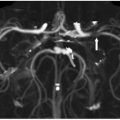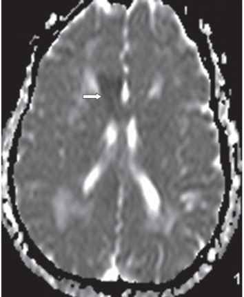
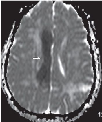
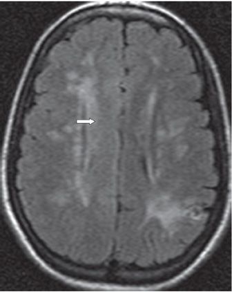
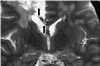
FINDINGS Figure 6-1. Axial DWI through the body of corpus callosum (CC). There is diffuse hyperintensity through the CC and cingulum on the right (arrow). Figures 6-2 and 6-3. ADC maps through the genu (Figure 6-2) and body (Figure 6-3) of the CC. There is low ADC in the genu and through the entire body of the CC and cingulum on the right (arrows). Figure 6-4. Axial T2 FLAIR through the body of CC. There is diffuse hyperintensity in the CC on the right (arrow). Multifocal bilateral white matter (WM) hyperintensities and a left parietal old infarct are also present. Figure 6-5. Coronal T2WI through the CC about 9 months later. There is atrophy and hyperintensity of the CC on the right side (arrows). Associated right parasagittal frontal lobe chronic infarct and widespread bilateral WM changes are present. There is ex vacuo dilatation of the right lateral ventricle.
DIFFERENTIAL DIAGNOSIS
Stay updated, free articles. Join our Telegram channel

Full access? Get Clinical Tree


