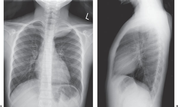
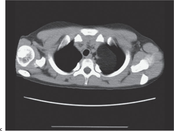
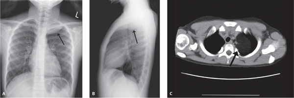
Case 6 A teenager with shortness of breath. (A,B) Frontal and lateral chest radiographs demonstrate a smooth, well-defined soft-tissue mass in the apex of the left hemithorax (arrows). (C) Axial post-contrast computed tomography (CT) image demonstrates that the smooth, homogeneous left apical mass has an attenuation value approximately equal to that of the subcutaneous adipose tissue (arrow).
Clinical Presentation
Further Work-up
Imaging Findings
Stay updated, free articles. Join our Telegram channel

Full access? Get Clinical Tree


