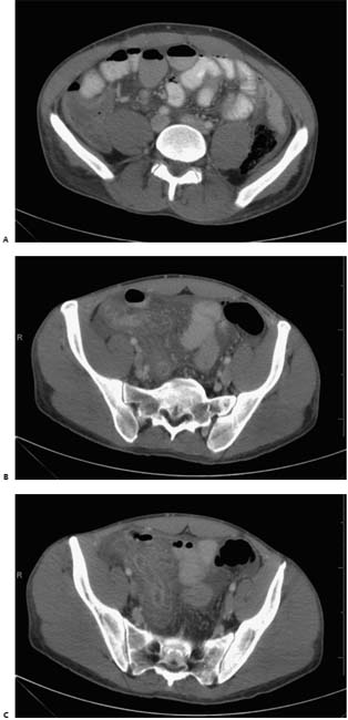
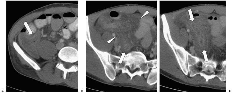
Case 6 A 45-year-old man presents with severe pelvic pain. (A) Infused abdominal computed tomography (CT) shows thickening of the cecal wall (arrow). (B) Lower image shows diffuse infiltration of the mesenteric fat (arrowheads) within the right lower quadrant (RLQ) and pelvis. A round, enhancing structure (arrow) is seen in the pelvis. (C) Lower image shows this structure to be tubular (arrows), with an enhancing wall and central fluid density. • Acute appendicitis: Acute appendicitis should be the first consideration for a tubular pelvic structure with a thick enhancing wall, mesenteric fat infiltration, and cecal wall thickening. • Crohn disease:
 Clinical Presentation
 Imaging Findings
 Differential Diagnosis
![]()
Stay updated, free articles. Join our Telegram channel

Full access? Get Clinical Tree


