Case 6
 Clinical Presentation
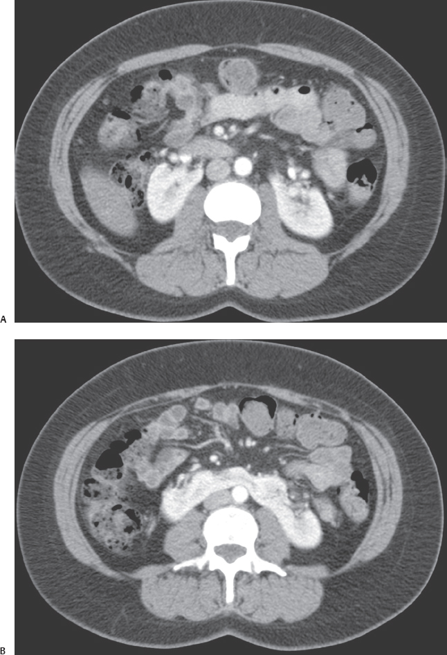
A 38-year-old woman who underwent contrast-enhanced computed tomography for vague abdominal pain.
 Imaging Findings
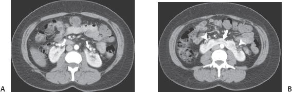
(A) Contrast-enhanced computed tomography (CT) image of the abdomen at the level of the kidneys obtained during the nephrographic phase shows that the kidneys are malrotated, with the renal hila pointing forward (arrows). No abnormality of size, shape, location, outline, or parenchymal thickness is present. (B) Contrast-enhanced CT images of the abdomen at a level lower than that of Figure A shows that the lower poles of both kidneys are joined by an isthmus, which is the bar of enhancing renal tissue crossing the spine (arrow). The ureters can be identified anterior to this isthmus (arrowheads).
 Differential Diagnosis
• Horseshoe kidney: The characteristic feature on CT is an isthmus that always connects the lower poles of both kidneys. It may vary from a thin fibrotic strand to full parenchymal thickness.
• Simple nonrotation: In one or both kidneys, the renal sinus may point anteriorly. However, there is no connecting isthmus.
Stay updated, free articles. Join our Telegram channel

Full access? Get Clinical Tree


