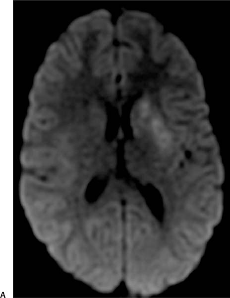
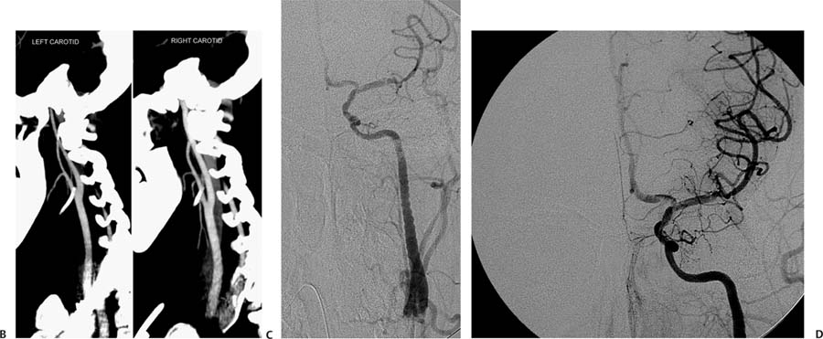
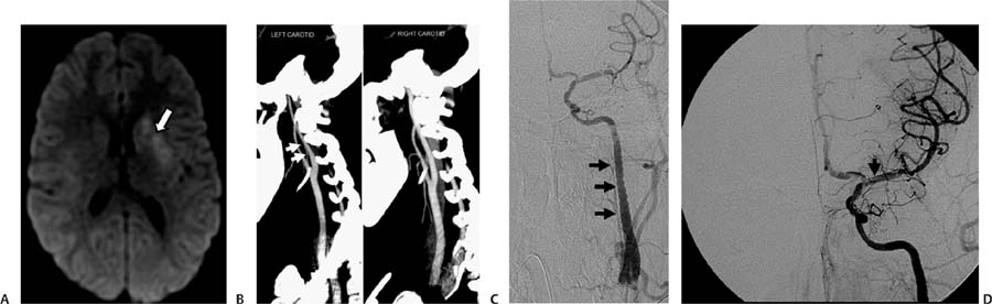
Case 60 A 17-year-old girl who fainted while playing basketball. (A) Diffusion-weighted image demonstrates acute ischemic changes in the left caudate head and putamen (arrow). (B) Sagittal computed tomography angiography maximum-intensity-projection images of the bifurcations of both carotid arteries demonstrate long-segment narrowing of the left internal carotid artery (ICA) with a serrated contour (arrows). (C) Lateral projection of a digital subtraction angiogram of the left ICA shows a “string of beads” appearance in the cervical segment (arrows). (D) Intracranial views of the same injection show a linear filling defect in the proximal left middle cerebral artery, consistent with a dissection flap (arrow). Irregular filling defects in the distal carotid artery and anterior cerebral artery are also noted, which represent thrombi.
Clinical Presentation
Further Work-up
Imaging Findings
Differential Diagnosis
Stay updated, free articles. Join our Telegram channel

Full access? Get Clinical Tree


