 Clinical Presentation
A 58-year-old man with hoarseness and cough.
Further Work-up
 Imaging Findings
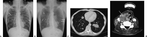
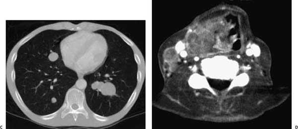
(A) Chest radiograph demonstrates multiple lower lung zone–predominant well-circumscribed pulmonary nodules (arrows). (B) Energy-subtracted chest radiograph demonstrates the pulmonary nodules to better advantage (arrows). (C) Contrast-enhanced computed tomography (CT; lung window) at the level of the coronary sinus confirms multiple well-circumscribed noncalcified pulmonary nodules (arrows). (D) Contrast-enhanced CT (soft-tissue window) at the level of the vocal cords demonstrates a large heterogeneous laryngeal mass (arrow) with right jugular lymphadenopathy.
 Differential Diagnosis
• Pulmonary metastases of head and neck carcinoma:
Stay updated, free articles. Join our Telegram channel

Full access? Get Clinical Tree


