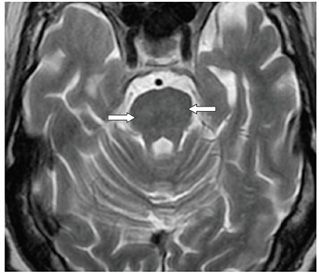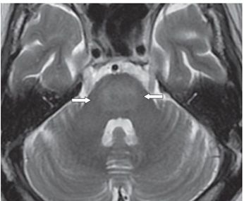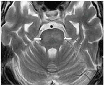


FINDINGS Figures 60-1 and 60-2. Two contiguous axial T2WI through the pons showing mild smudgy hyperintensity in the basis pontis. Lesion is very subtle (arrows). Figures 60-3 and 60-4. Follow-up images 8 days later reveal a rather prominent round central pontine hyperintensity with fluffy outline and peripheral sparing (arrows). There were no other significant abnormalities elsewhere other than mild white matter changes and volume loss. There was no restricted diffusion or abnormal contrast enhancement.
DIFFERENTIAL DIAGNOSIS Osmotic demyelination syndrome (ODS)/central pontine myelinolysis (CPM), chronic small vessel ischemic disease, encephalitis, brainstem posterior reversible encephalopathy syndrome (PRES).
DIAGNOSIS Osmotic demyelination syndrome (ODS)/central pontine myelinolysis (CPM).
DISCUSSION
Stay updated, free articles. Join our Telegram channel

Full access? Get Clinical Tree


