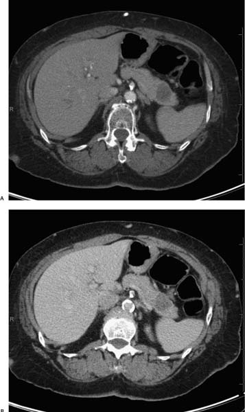
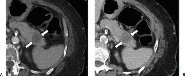
Case 60 A 61-year-old man presents with chronic back pain. (A) Contrast-enhanced computed tomography (CT) in the arterial phase shows a well-circumscribed, hypodense mass (arrows) with punctate calcifications in the tail of the pancreas, without associated lymphadenopathy or infiltration of adjacent structures. (B) Venous-phase image shows a moderately enhancing mass (arrows) with a granular appearance suggesting multiple small cysts, internal septations, and the hint of a central scar. • Microcystic adenoma: This is the most likely diagnosis, suggested by the subtle appearance of multiple small cysts, septa, and a central scar. A demonstration of stability on serial CT scans would further suggest this benign entity. • Pancreatic adenocarcinoma:
 Clinical Presentation
 Imaging Findings
 Differential Diagnosis
![]()
Stay updated, free articles. Join our Telegram channel

Full access? Get Clinical Tree


