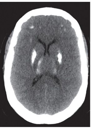
FINDINGS Figure 61-1. Axial NCCT through the basal ganglia. There are significant bilateral almost symmetrical calcifications in the basal ganglia, thalami, frontal white matter, and occipital cortex. There is no atrophy of the brain. Figure 61-2. Axial NCCT in a different patient. There is lesser degree of bilateral symmetrical calcifications in the basal ganglia, thalami, and subcortical frontal white matter.
DIFFERENTIAL DIAGNOSIS Hypoparathyroidism, pseudohypoparathryroidism, pseudopseudohypoparathyroidism, hypothyroidism, physiologic calcifications, Fahr disease (bilateral striopallidodentate calcinosis).
DIAGNOSIS Fahr disease (bilateral striopallidodentate calcinosis).
DISCUSSION
Stay updated, free articles. Join our Telegram channel

Full access? Get Clinical Tree


