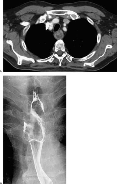
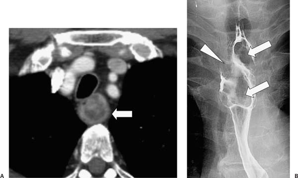
Case 61 A 62-year-old man presents with dysphagia. (A) Contrast-enhanced computed tomography (CT) shows an upper esophageal soft-tissue mass (arrow) with a necrotic center. (B) Single-contrast esophagogram shows a large, intraluminal, multilobular polypoid mass (arrows). The upper right side of the mass shows a broad, pedunculated attachment to the esophageal wall (arrowhead). • Spindle cell carcinoma: This is the most likely diagnosis. It is characteristically bulky and polypoid and expands the lumen without complete obstruction, which is consistent with the appearance of this case on the esophagogram.
 Clinical Presentation
 Imaging Findings
 Differential Diagnosis
Stay updated, free articles. Join our Telegram channel

Full access? Get Clinical Tree


