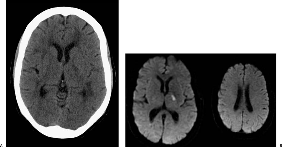
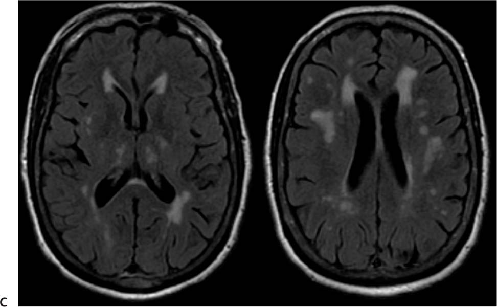
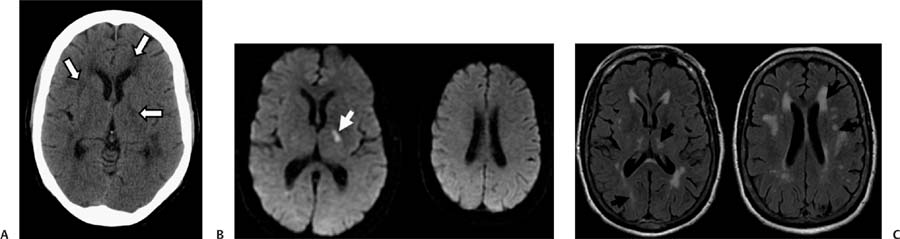
Case 62 A 47-year-old woman with transient weakness. (A) Computed tomography (CT) of the brain shows numerous small areas of low attenuation in the deep white matter bilaterally and in the left thalamus (arrows). (B) Diffusion-weighted image shows restriction in the left thalamus (arrow). The centrum semiovale is unremarkable. (C) Fluid-attenuated inversion recovery (FLAIR) images demonstrate numerous areas of high signal (arrows), including the lesion with restricted diffusion in the left thalamus. • Acute and chronic lacunar infarcts:
Clinical Presentation
Further Work-up
Imaging Findings
Differential Diagnosis
![]()
Stay updated, free articles. Join our Telegram channel

Full access? Get Clinical Tree


