 Clinical Presentation
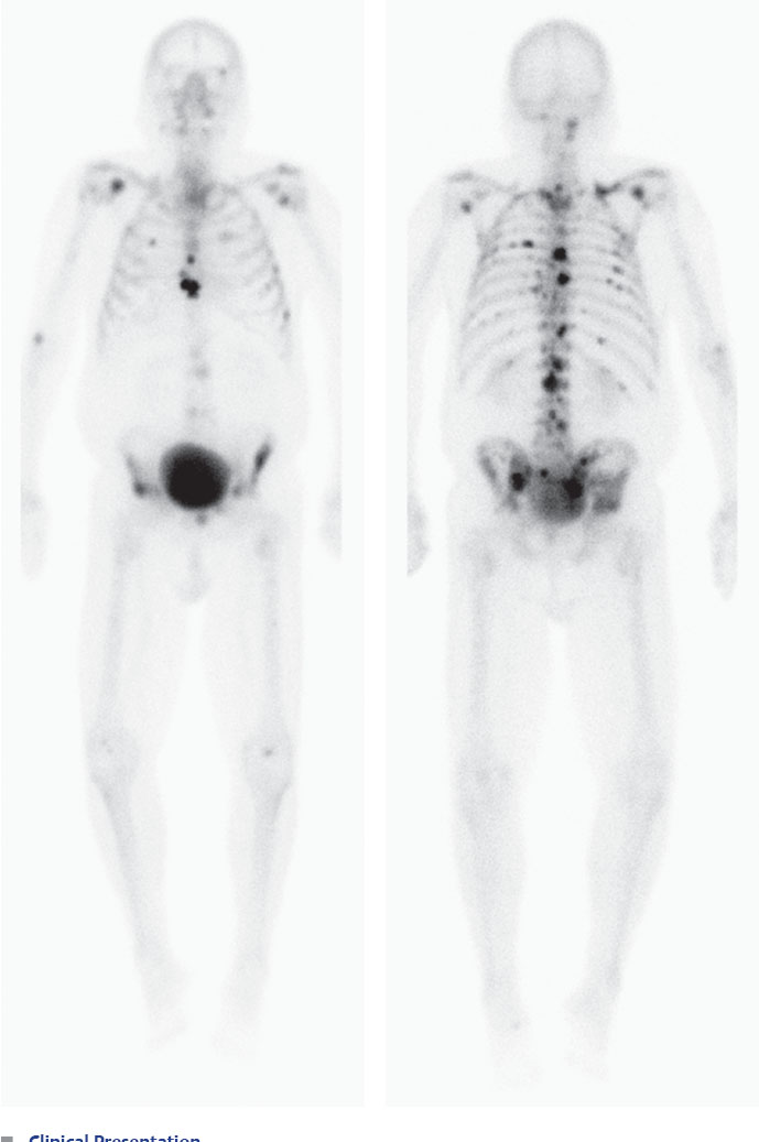
A 57-year-old man with a history of prostate cancer and diffuse body pain.

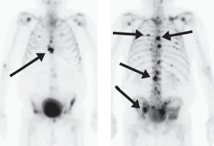
Three-hour–delayed anterior and posterior whole-body bone scan demonstrates too-numerous-to-count osteoblastic lesions in the axial and proximal appendicular skeleton (arrows).
 Differential Diagnosis
• Diffuse osteoblastic metastases: This pattern of uptake is most consistent with diffuse osseous metastatic disease. Note that there is minimal abnormal uptake in the appendicular skeleton (beyond red marrow distribution).
• Multifocal Paget disease: This can also demonstrate multiple lesions. However, these need not be confined to the axial skeleton and typically appear as long-segment lesions that, in the extremities, start at the bone ends (see Case 17).
• Multiple bone lesions associated with a metabolic disorder: Occasionally, hyperparathyroidism or other metabolic conditions can give the appearance of multiple bony lesions that can be apparent on bone scan. However, the lack of significant uptake in the peripheral appendicular skeleton argues against this possibility.

Related posts:


Stay updated, free articles. Join our Telegram channel

Full access? Get Clinical Tree


