 Clinical Presentation
A 50-year-old woman with dyspnea on exertion.
Further Work-up

 Imaging Findings
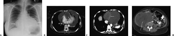
(A) Chest radiograph demonstrates a globular enlarged cardiac silhouette (arrows). Linear atelectasis is seen in the left lung. (B) Contrast-enhanced computed tomography (CT) through the right ventricle (soft-tissue window) shows a large pericardial effusion. A large mass is invading the right ventricle (arrow). (C) Contrast-enhanced CT through the caudal margin of the pericardium (soft-tissue window) shows an extensive amount of pericardial fluid. Although the fluid is predominantly of low density, a curvilinear area of high density is seen anterior to the inferior vena cava (IVC, arrow). (D) Contrast-enhanced CT (soft-tissue window) through the abdomen demonstrates a large mass occupying most of the right abdomen (long arrow). There is a resultant mass effect on the ascending colon and IVC. The density is heterogeneous, but there are extensive areas of very low density.
 Differential Diagnosis
Stay updated, free articles. Join our Telegram channel

Full access? Get Clinical Tree


