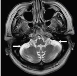
FINDINGS Figures 62-1 and 62-2. Sagittal T1WI and axial T2WI through the cerebellum respectively. There is prominence of the pericerebellar cerebrospinal fluid (CSF) spaces with significant thinning of the cerebellar folia and widening of intervening CSF spaces—fissures (arrow). There is normal appearing brainstem. The remainder of the brain (partially shown in Figure 62-1) is relatively normal in morphology.
DIFFERENTIAL DIAGNOSIS Normal aging, drug-related atrophy, alcohol-related atrophy, cerebellar ataxia syndrome, paraneoplastic disease.
DIAGNOSIS Cerebellar ataxia syndrome (inherited).
DISCUSSION
Stay updated, free articles. Join our Telegram channel

Full access? Get Clinical Tree


