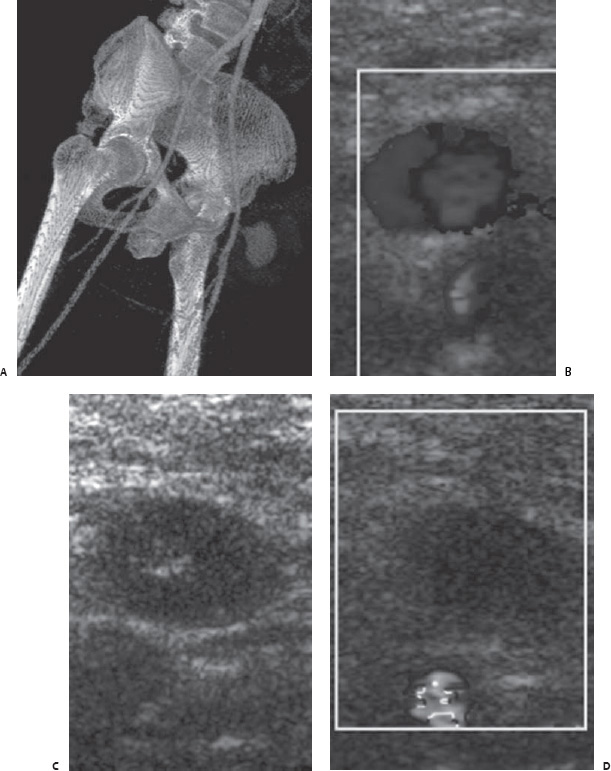
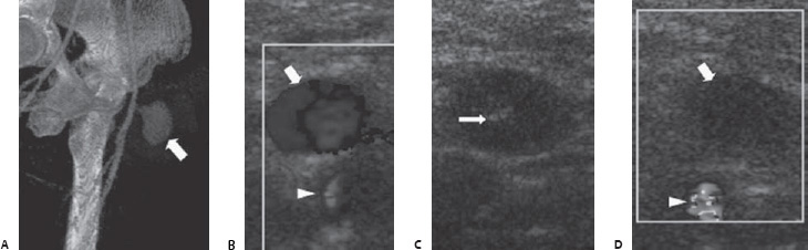
Case 62 A 51-year-old man presents with swelling in the left groin 2 days after catheterization of the left common femoral artery. (A) Computed tomographic angiography (CTA) shows a pseudoaneurysm of the left common femoral artery (CFA: arrow). (B) Color Doppler shows “yin-yang” appearance of the pseudoaneurysm (arrow). The common femoral artery is also shown (arrowhead). (C) Grayscale image shows a 22-gauge needle in the center of the pseudoaneurysm (arrow). (D) Color Doppler after thrombin injection shows cessation of flow in the pseudoaneurysm (arrow). The common femoral artery retains flow (arrowhead).
 Clinical Presentation
 Imaging Findings
 Differential Diagnosis
 Essential Facts
Stay updated, free articles. Join our Telegram channel

Full access? Get Clinical Tree


