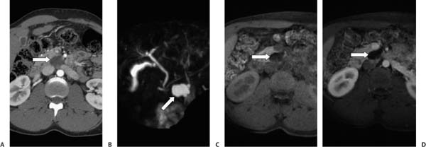
Case 62 A 66-year-old man presents with back pain, diarrhea, and fever. (A) Contrast-enhanced computed tomography (CT) shows a low-density, well-circumscribed mass (arrow) in the uncinate process of the pancreas. (B) Magnetic resonance cholangiopancreatography (MRCP) shows the mass (arrow) to be communicating with the ventral pancreatic duct and hyperintense on T2. (C) Preinfusion T1 image shows the mass (arrow) to be hypointense. (D) Postinfusion T1 image shows no enhancement (arrow). • Intraductal papillary mucinous neoplasm (IPMN): This is the first choice for the diagnosis. It most often occurs in men and has a cystic component communicating with a pancreatic duct. The focal type is more common in the pancreatic head. It is distinguished by direct communication of the cyst with the main pancreatic duct or a side branch. • Pancreatic pseudocyst:
 Clinical Presentation
 Imaging Findings
 Differential Diagnosis
![]()
Stay updated, free articles. Join our Telegram channel

Full access? Get Clinical Tree


