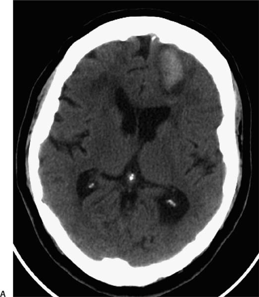
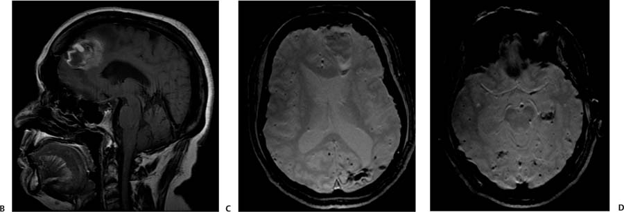
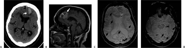
Case 63 A 78-year-old man with a history of prior cerebral hemorrhage presenting with headache and right leg weakness. (A) Nonenhanced computed tomography (CT) of the brain demonstrates acute hemorrhage in the left frontal lobe (arrow). (B) Sagittal T1-weighted image (WI) of the brain shows a subacute hematoma in the right frontal lobe (arrow). (C) Gradient-echo (GRE) T2*WI shows an acute left frontal hematoma, an old left occipital hemorrhagic lesion, and multiple punctate foci of hemosiderin deposition in the subcortical regions of both cerebral hemispheres (arrows). (D) GRE T2*WI: note the multiple punctate foci of hemosiderin deposition in both cerebral hemispheres (arrows).
Clinical Presentation
Further Work-up
Imaging Findings
Differential Diagnosis
Stay updated, free articles. Join our Telegram channel

Full access? Get Clinical Tree


