 Clinical Presentation
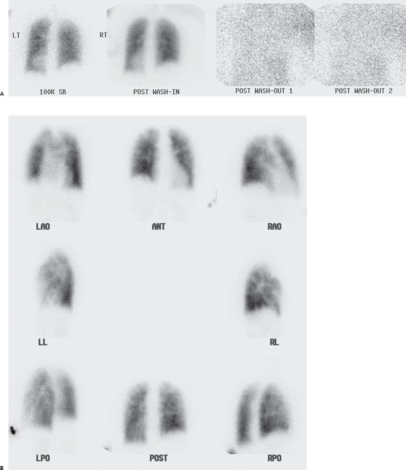
A 64-year-old woman with breast cancer presents with shortness of breath.

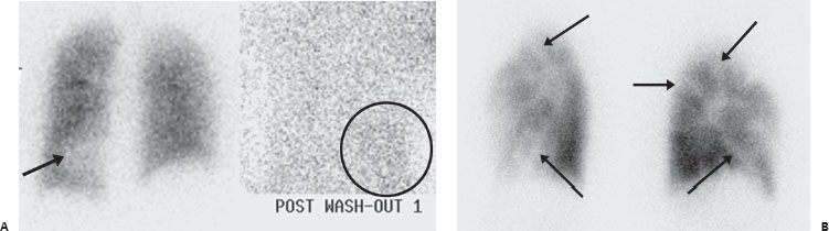
(A) Sequential Xenon 133 ventilation images demonstrate focal decrease in single-breath ventilation at the left lung base (arrow), but with the remaining distribution uniform. There is no abnormal retention. Faint accumulation of tracer is seen on washout in a fatty liver (circle) below an elevated right hemidiaphragm (see Case 9). (B) Tc99m-MAA images demonstrate multiple linear perfusion defects outlining the periphery of most segments: the “segmental contouring” sign or “contour mapping” sign (arrows).
 Differential Diagnosis
• Diffuse tumor microemboli (TM): Multiple linear mismatched perfusion defects outlining the periphery of most segments make this the correct diagnosis.
Related posts:


Stay updated, free articles. Join our Telegram channel

Full access? Get Clinical Tree


