 Clinical Presentation
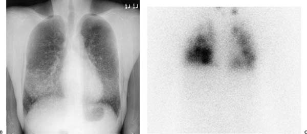
A 25-year-old woman with dyspnea.
Further Work-up

 Imaging Findings

(A) Frontal chest radiograph demonstrates innumerable small, high-density pulmonary nodules that are lower lobe–predominant (arrows). Given their density, many of the nodules are likely calcified. (B) Energy-subtracted frontal chest radiograph demonstrates the nodules (arrows) to better advantage. (C) Planar anterior thoracic image from an iodine 131 (131I) whole-body scan demonstrates intense lower lobe–predominant uptake (arrows). Uptake is also seen within the neck.


