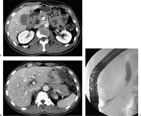
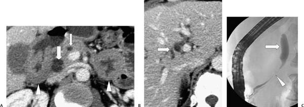
Case 63 A 58-year-old man presents with weight loss and mild right upper quadrant abdominal pain. (A) Contrast-enhanced computed tomography (CT) shows marked, diffuse thickening of the duodenal wall (arrowheads), which is inseparable from the pancreatic head, and dilatation of both the common bile duct (large arrow) and the pancreatic duct (small arrow). (B) Higher cut shows intrahepatic biliary dilatation (arrow). (C) Endoscopic retrograde cholangiopancreatography shows dilatation of the common bile duct (large arrow) with eccentric, shelflike transition to a long, distal stricture (arrowhead). • Duodenal lymphoma: This infiltrating mass affecting a long segment of small bowel is more likely to be lymphoma than infiltrating adenocarcinoma; hence, it is the first choice for the correct diagnosis. • Metastatic disease:
 Clinical Presentation
 Imaging Findings
 Differential Diagnosis
![]()
Stay updated, free articles. Join our Telegram channel

Full access? Get Clinical Tree


