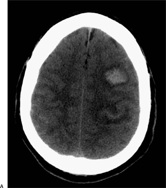
Case 64 A 72-year-old man with a history of treated cancer, now presenting with severe headache. (A) Axial computed tomography (CT) of the brain demonstrates subarachnoid hemorrhage at the vertex bilaterally (arrows) and parenchymal hematoma in the left frontal lobe. (B) Axial T1-weighted image (WI) shows increased signal in the superior longitudinal sinus (arrow). The left frontal hematoma is isointense to the cortex (acute). (C) Coronal T2WI shows subdural hemorrhage in the parafalcine region (arrowhead). The parenchymal hematoma shows a central area of low signal and a rim of high signal. (D) Lateral projection digital subtraction angiography of the venous phase of the left carotid injection demonstrates multiple filling defects in the sagittal sinus (arrows). • Parenchymal and extra-axial hemorrhages secondary to venous sinus thrombosis:
Clinical Presentation
Further Work-up
Imaging Findings
Differential Diagnosis
![]()
Stay updated, free articles. Join our Telegram channel

Full access? Get Clinical Tree


