 Clinical Presentation
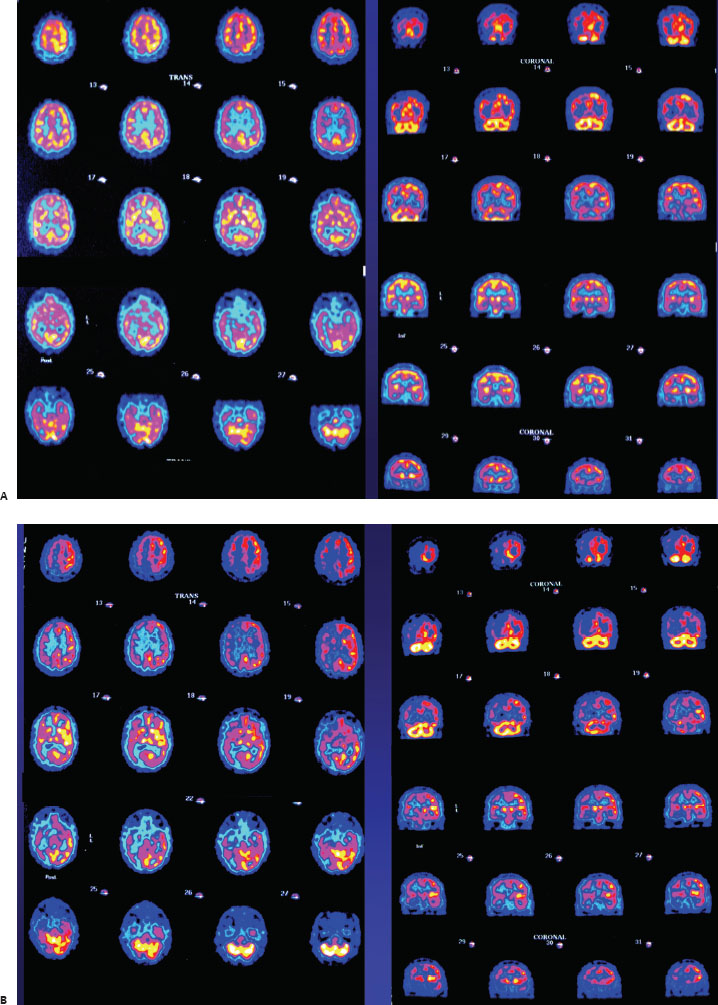
A 64-year-old man with transient ischemic attacks. The exam was acquired before (A) and after (B) a pharmacologic intervention.

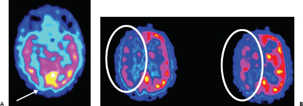
(A) Axial and coronal brain perfusion SPECT images obtained with Tc99m-HMPAO demonstrate a small cortical defect in the right posterior temporal–occipital location (arrow). The remainder of the perfusion is normal. (B) Axial and coronal brain perfusion SPECT images following acetazolamide administration demonstrate markedly decreased perfusion to most of the right cerebral hemisphere (circles).
 Differential Diagnosis
• Diminished cerebral vascular blood flow reserve:


Stay updated, free articles. Join our Telegram channel

Full access? Get Clinical Tree


