 Clinical Presentation
A 30-year-old woman with fatigue.
Further Work-up
 Imaging Findings
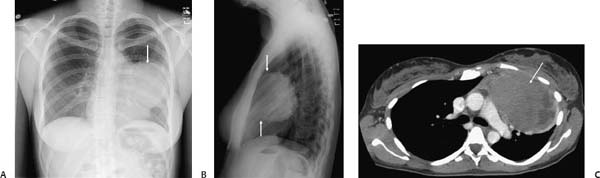
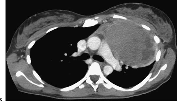
(A) Frontal chest radiograph demonstrates a large mass in the medial left hemithorax (arrow). There is loss of the left border of the heart, but the left hilar structures project through the mass (hilum overlay sign). (B) Lateral chest radiograph confirms the anterior location of the mass (arrows). (C) Contrast-enhanced computed tomography (CT; soft-tissue window) demonstrates a heterogeneous anterior mediastinal mass (arrow) that abuts the thymus.
 Differential Diagnosis
• Lymphoma: A solitary noncalcified anterior mediastinal mass separate from the thymus in a patient of this age is suggestive of lymphoma. Although this patient had large B-cell non-Hodgkin lymphoma, Hodgkin disease (HD) would be the most likely diagnosis because most mediastinal lymphoma is HD.
• Teratoma:
Stay updated, free articles. Join our Telegram channel

Full access? Get Clinical Tree


