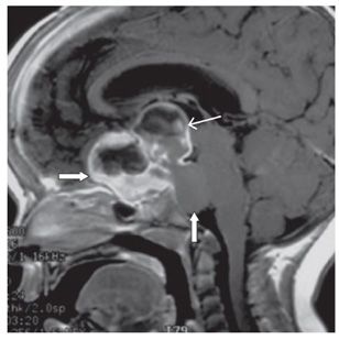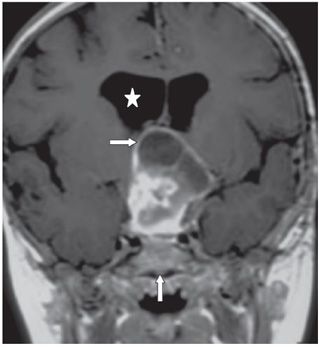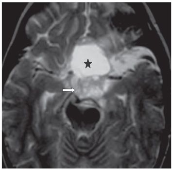


FINDINGS Figure 64-1. Sagittal non-contrast T1WI. There is a large multilobulated solid cystic mass in the suprasellar cistern. There is marked mass effect and displacement of the floor of the third ventricle (vertical arrow), posterior inferior frontal lobes (transverse arrow), midbrain and pons (star). The lesion is heterogeneous hypo- to isointense. Figure 64-2. Sagittal post-contrast T1WI. Thin (thin arrow) and thick (thick anterior arrow) walls of enhancement are present. The component in front of the pons (vertical arrow) is not enhancing. Figure 64-3. Coronal post-contrast T1WI. The superior extension of the lesion to the foramina of Monro (transverse arrow) and the dilation of the lateral ventricles (star) are demonstrated. The sella turcica is compressed (vertical arrow). Figure 64-4. Axial T2WI. The cystic component is noted to be hyperintense to cerebrospinal fluid (CSF) (star), and the more solid component is only mildly hyperintense (arrow). There is mild surrounding edema. No evidence of calcifications was found on the MRI.
DIFFERENTIAL DIAGNOSIS Optic nerve glioma, craniopharyngioma, meningioma, metastatic tumor, macroadenoma, aneurysm, sarcoid, tuberculoma, pilocytic astrocytoma (PA).
DIAGNOSIS PA of optic pathway.
DISCUSSION
Stay updated, free articles. Join our Telegram channel

Full access? Get Clinical Tree


