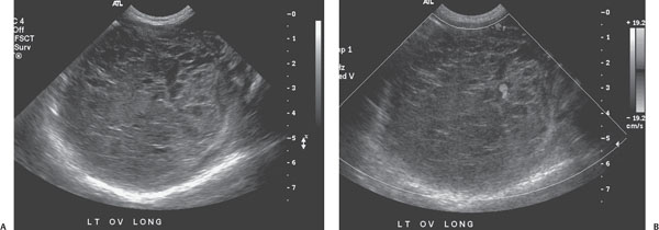
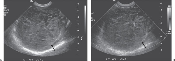
Case 64 A neonatal girl with an abdominal mass. (A,B) Longitudinal gray-scale and Doppler sonograms of the pelvis demonstrate a markedly enlarged (> 6 cm in diameter), heterogeneously hypoechoic left ovary containing multiple peripheral cysts (arrows). The ovary shows almost no internal Doppler signal. • Ovarian torsion:
Clinical Presentation
Imaging Findings
Differential Diagnosis
![]()
Stay updated, free articles. Join our Telegram channel

Full access? Get Clinical Tree


