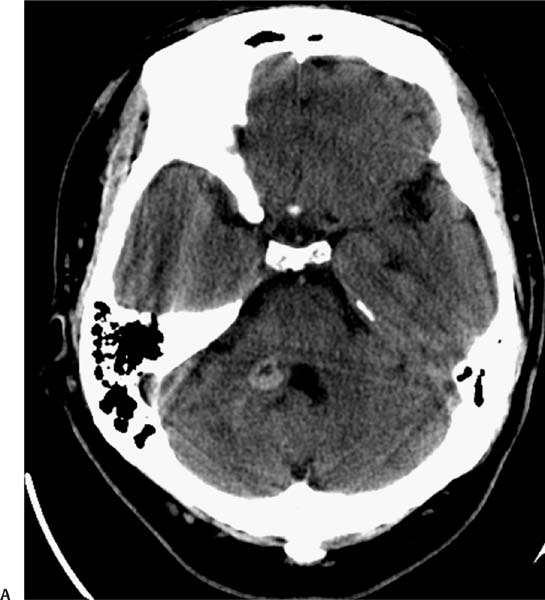
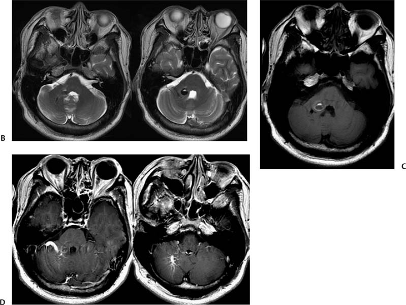
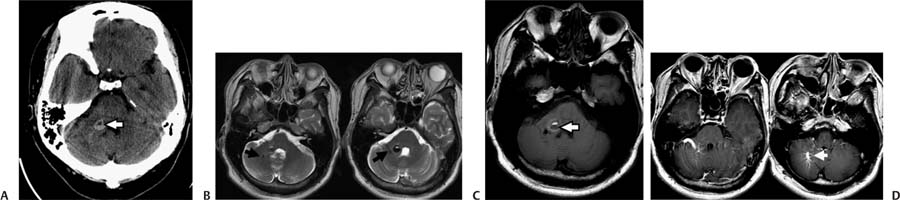
Case 65 A 35-year-old with the new onset of seizures. (A) Nonenhanced computed tomography (CT) demonstrates a round area of increased attenuation with a fluidlike core in the right middle cerebellar peduncle (arrow). There is no edema or mass effect. (B) Axial T2-weighted images (WIs) show a round lesion with heterogeneous intensity. A rim of low signal is evident. There are a few linear flow voids lateral to this lesion (arrows). (C) The areas of high signal on T2WIs are also hyperintense on T1WIs. The dark rim on T2WIs has intermediate signal on T1WIs. Findings are consistent with blood products of different ages (arrow). (D) Axial T1WIs after contrast show enhancement of vascular structures, with a linear and a “caput medusae” pattern (arrow). • Cavernous malformation (CM) with associated developmental venous anomaly (DVA):
Clinical Presentation
Further Work-up
Imaging Findings
Differential Diagnosis
![]()
Stay updated, free articles. Join our Telegram channel

Full access? Get Clinical Tree


