 Clinical Presentation
A 32-year-old woman with chest pain.
Further Work-up
 Imaging Findings
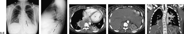
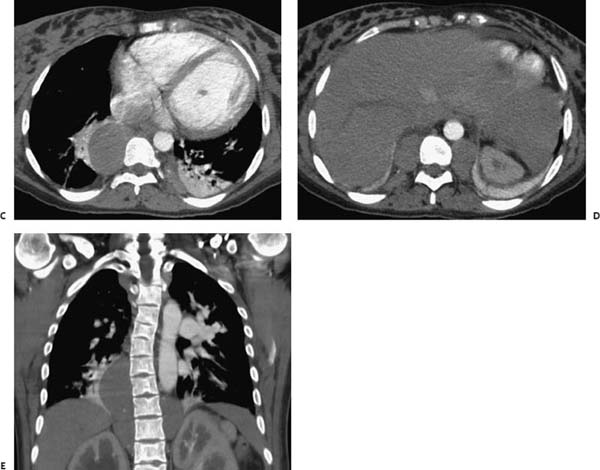
(A) Frontal chest radiograph demonstrates a right paraspinal mass with abnormal convexity of the upper azygoesophageal and right paraspinal lines (white arrow). The left paraspinal line is also effaced, although the lateral margin of the aorta is still seen (black arrow). There is mild cardiomegaly and basilar atelectasis. (B) Lateral chest radiograph confirms the posterior location of the mass (arrows). There is also multilevel vertebral end plate depression, creating “H-type” vertebrae. (C) Contrast-enhanced computed tomography (CT; soft-tissue window) through the level of the heart confirms a right paraspinal mass with adjacent atelectasis (arrow). The mass has homogeneously low attenuation. There is no evidence of osseous erosion. A much smaller lesion is seen on the left. (D) Contrast-enhanced CT (soft-tissue window) through the upper abdomen shows bilateral hypodense paraspinal masses (arrows). The lesions have smooth borders, and there is no associated osseous erosion. Hepatomegaly is present, but the spleen is not seen. (E) Coronal CT shows the paraspinal masses (white arrows) and vertebral body abnormalities (black arrows) to better advantage. There is also sclerosis of the left humeral head.
Stay updated, free articles. Join our Telegram channel

Full access? Get Clinical Tree


