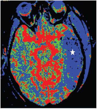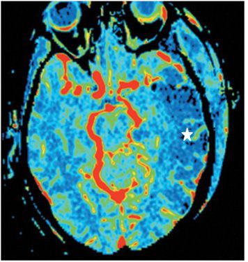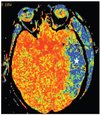


FINDINGS Figure 65-1. Axial NCCT through the temporal lobes. There is a left temporal lobe diffuse hypodensity (star) with mass effect and subtle left uncal herniation consistent with malignant acute ischemic infarct. There is a left temporal scalp soft tissue swelling from recent surgery (arrow). Figure 65-2. Axial CT perfusion (CTP) blood flow color map through the temporal lobes. There is significantly diminished blood flow in the left temporal lobe (star). Figure 65-3. Axial CTP blood volume color map through the temporal lobes. There is diminished left temporal lobe blood volume (star) of same size as the flow deficit. Figure 65-4. CTP mean transit time (MTT) color map through the temporal lobes. There is prolonged MTT (star) of similar size to the blood flow and volume deficits. These are matching perfusion maps consistent with infarction without significant area of salvageable penumbra.
DIFFERENTIAL DIAGNOSIS N/A.
DIAGNOSIS CTP of left MCA territory ischemic infarct.
DISCUSSION
Stay updated, free articles. Join our Telegram channel

Full access? Get Clinical Tree


