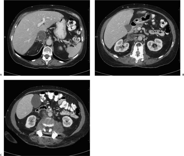
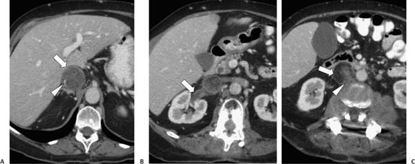
Case 65 A 50-year-old woman presents with back pain, nausea, and weight loss. (A) Contrast-enhanced computed tomography (CT) shows marked enlargement of the inferior vena cava (IVC; arrow) with obliteration of the lumen by soft tissue. Arterial branches (arrowhead) course through this soft tissue. (B) More caudal image shows extension of the mass into the right renal vein (arrow). The mass appears more heterogeneous in density at this level. The kidney parenchyma is normal. (C) More caudal image shows a fatty component (arrow) of the intraluminal IVC mass, which extends outside the confines of the IVC into the retroperitoneal fat (arrowhead). • Liposarcoma locally invasive to the IVC: This is the most likely explanation for a vascular soft-tissue mass with a component of macroscopic fat occurring within the retroperitoneum, albeit within the IVC itself.

 Clinical Presentation
 Imaging Findings

 Differential Diagnosis
Stay updated, free articles. Join our Telegram channel

Full access? Get Clinical Tree


