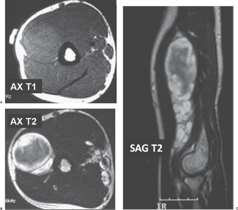
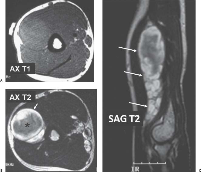
Case 66 A 30-year-old man presents with scattered areas of skin discoloration and soft-tissue masses along the right arm. (A–C) Magnetic resonance (MR) images of the right arm reveal convoluted multinodular masses and thickening (long arrows) along the expected course of the radial nerve. The axial T2-weighted MR image shows high signal intensity peripherally (short arrow) with low signal intensity centrally (asterisk), the “target sign.”

 Clinical Presentation
 Imaging Findings

Stay updated, free articles. Join our Telegram channel

Full access? Get Clinical Tree


