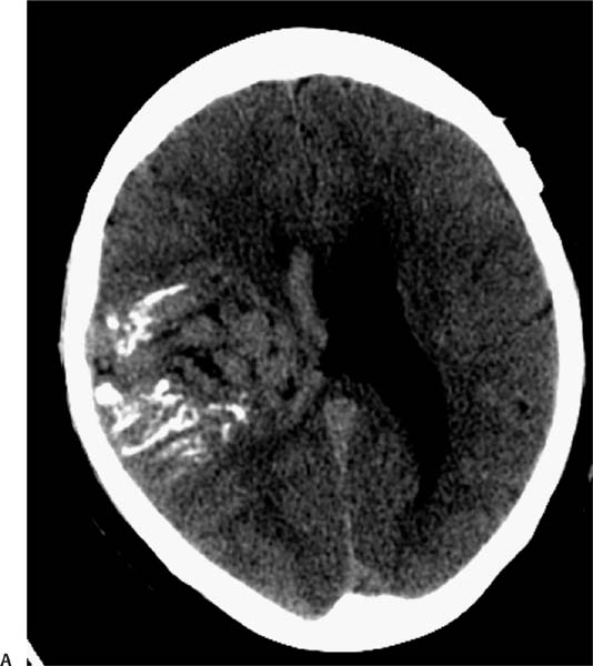
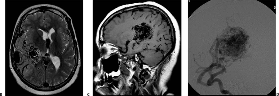
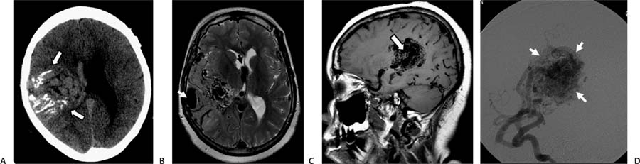
Case 66 A 55-year-old woman with headaches and left hemiparesis. (A) Computed tomography (CT) of the brain shows a large right hemispheric lesion with linear calcifications and serpentine areas of increased attenuation (arrows). Note that the mass effect of the lesion is small relative to its size. (B) Axial T2-weighted image (WI) shows numerous large flow voids surrounded by areas of high signal in the temporal and parietal lobes, indicative of gliosis. The internal cerebral veins (black arrow) and superficial veins (white arrow) are enlarged. (C) Sagittal T1WI shows flow voids in a large arteriovenous nidus (arrow). (D) Right internal carotid angiogram shows early filling of a large arteriovenous nidus (arrows). • Arteriovenous malformation (AVM):
Clinical Presentation
Further Work-up
Imaging Findings
Differential Diagnosis
![]()
Stay updated, free articles. Join our Telegram channel

Full access? Get Clinical Tree


