 Clinical Presentation
A 30-year-old man with respiratory distress.
Further Work-up
 Imaging Findings
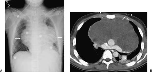
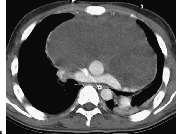
(A) Chest radiograph demonstrates an intubated patient with a large chest mass (arrows). Its borders with the cardiomediastinal silhouette suggest an anterior mediastinal location. There are no vertebral body or rib abnormalities. (B) Contrast-enhanced computed tomography of the chest (soft-tissue windows) confirms that the mass is located in the anterior mediastinum (arrow
Stay updated, free articles. Join our Telegram channel

Full access? Get Clinical Tree


