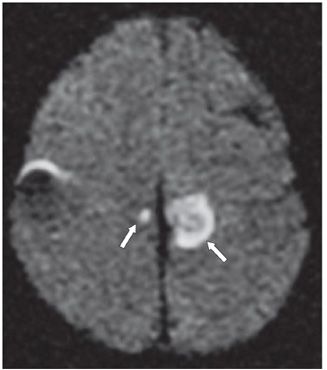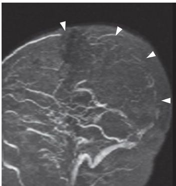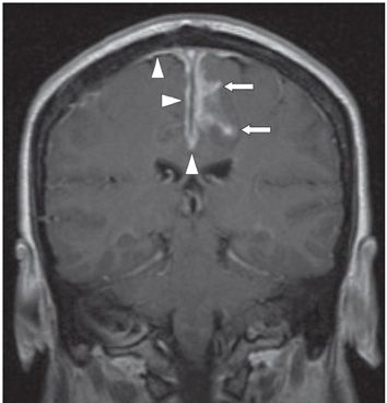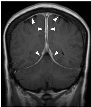


FINDINGS Figure 66-1. Axial FLAIR image through the centrum semiovale. There is bilateral subcortical white matter signal hyperintensity within the centrum semiovale (arrowheads). There is a suggestion of increased signal intensity in the posterior aspect of the sagittal sinus. There is also focally hyperintense FLAIR signal involving the cortex of the medial surface of the left hemisphere (paracentral lobule*) that shows corresponding diffusion restriction on axial DWI in Figure 66-2 (arrows). Note the small, medial contralateral focus of diffusion restriction (Note: the DWI signal hyperintensity along the right convexity is artifactual). Figure 66-3. Sagittal 2D TOF MR venography MIP image shows absence of flow-related enhancement along the mid and posterior aspect of the superior sagittal sinus (arrowheads). Figures 66-4 and 66-5. Coronal post-contrast T1WI anteriorly and posteriorly, respectively. There is abnormal pachymeningeal thickening and enhancement (small arrowheads) in addition to sulcal leptomeningeal (pia-arachnoid) enhancement along the medial surface of the left cerebral hemisphere (small arrows).
DIFFERENTIAL DIAGNOSIS Dural and leptomeningeal metatastasis (breast, lymphoma), leukemia, en plaque and lymphoplasmacyte-rich meningiomas (LRM), neurosarcoidosis, Wegener granulomatosis, tuberculous meningitis, noninfectious inflammatory meningitis (i.e., rheumatoid pachymeningitis), bacterial and fungal meningitis, hypertrophic pachymeningitis, idiopathic granulomatous meningitis, intracranial hypotension.
DIAGNOSIS Idiopathic granulomatous pachymeningitis complicated by secondary sagittal sinus thrombosis, venous congestion, and venous infarction.
DISCUSSION
Stay updated, free articles. Join our Telegram channel

Full access? Get Clinical Tree


