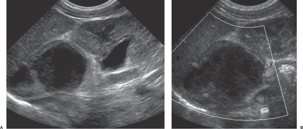
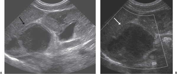
Case 66 A newborn with prenatal hydronephrosis. (A) Longitudinal gray-scale ultrasound (US) image: superior to the normal-appearing kidney is a large, predominately hypoechoic mass with a peripheral hyperechoic rim and internal debris (arrow). (B) Doppler US image: there is no blood flow within the lesion (arrow). • Adrenal hemorrhage: An avascular mass in the adrenal bed with a mixed echotexture is consistent with hemorrhage. • Neuroblastoma: Doppler US should show internal blood flow. • Congenital adrenal hyperplasia:
Clinical Presentation
Imaging Findings
Differential Diagnosis
![]()
Stay updated, free articles. Join our Telegram channel

Full access? Get Clinical Tree


