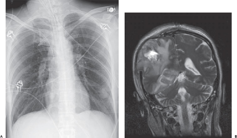
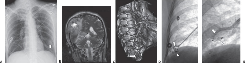
Case 66 A 37-year-old woman presents with hemiplegia. (A) Chest radiograph shows a well-circumscribed mass in the left lower lobe (arrow). (B) Magnetic resonance image shows a ring-enhancing lesion in the right hemisphere with mass effect. (C) Follow-up computed tomographic angiogram (CTA) shows an arteriovenous (a-v) malformation in the right lower lobe (arrow). Other a-v malformations were present bilaterally. (D) Selected pulmonary arteriogram shows artery (arrow) and vein (arrowhead) of the a-v malformation. (E) Repeat angiogram shows stasis in artery (arrow) supplying angiogram after deployment of an occluding device (arrowhead).
 Clinical Presentation
 Imaging Findings
 Differential Diagnosis
 Essential Facts
Stay updated, free articles. Join our Telegram channel

Full access? Get Clinical Tree


