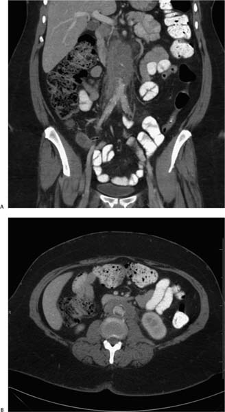
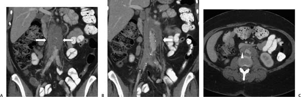
Case 66 A 69-year-old woman presents with flank pain. (A) Coronal reformatted infused computed tomography (CT) image shows a soft-tissue mass (arrows) encasing the inferior vena cava and aorta. (B) Adjacent image shows the mass encasing the left ureter (arrow). (C) Axial image shows the mass (arrowhead) enveloping the aorta and displacing the inferior vena cava. No vascular invasion is apparent. • Retroperitoneal fibrosis: This is the most likely diagnosis for soft-tissue infiltration of the retroperitoneum encasing, but not invading or displacing, retroperitoneal structures. • Retroperitoneal soft-tissue tumors:
 Clinical Presentation
 Imaging Findings
 Differential Diagnosis
![]()
Stay updated, free articles. Join our Telegram channel

Full access? Get Clinical Tree


